If your in vitro transcribed mRNA is producing lower yields, inconsistent results between batches, or immune activation you didn’t design for, there’s a reasonable chance you’ve already ruled out the obvious causes. Reagent lot, transfection efficiency, cell health, construct design — most labs work through that list before they get anywhere near the IVT reaction itself. What often gets missed is that the IVT reaction is where the problem started.
Every T7 RNA polymerase reaction produces double-stranded RNA as a byproduct. It isn’t a failure mode tied to a particular protocol or nucleotide choice — it’s a structural feature of how T7 RNA polymerase works. Snap-back synthesis at the 3′ end of a transcript, antisense priming from polymerase runoff past a linearized template, and abortive transcripts that re-prime a second round of synthesis all generate dsRNA species alongside your intended product. Canonical nucleotides, modified nucleotides, none of them prevent dsRNA formation, because none of them touch the mechanism that creates it.
What dsRNA actually does
Cells have a set of pattern recognition receptors — RIG-I, MDA5, TLR3, OAS1, PKR among them — that evolved specifically to detect double-stranded RNA, because dsRNA is a hallmark of viral replication intermediates. To these sensors, dsRNA carryover looks the same as an active infection. Once triggered, the response is what you’d expect from an antiviral program: type I interferon induction, translational shutdown, and inflammatory signaling that, when dsRNA levels are high, can compromise cell viability.
What tends to surprise people is how little dsRNA it takes. Even in nucleoside-modified mRNA, the residual dsRNA left after transcription is enough to drive type I interferon and inflammatory cytokine induction, and clearing it can raise protein expression by orders of magnitude in primary cells (1).
Modified nucleotides help. They don’t finish the job.
N1-methyl-pseudouridine and pseudouridine substitution are now standard practice for reducing mRNA immunogenicity, and for good reason — they measurably reduce activation of nucleoside-sensing pathways. What they don’t do is stop dsRNA from forming during transcription, because dsRNA activates a parallel set of sensors that respond to duplex structure rather than nucleoside chemistry.
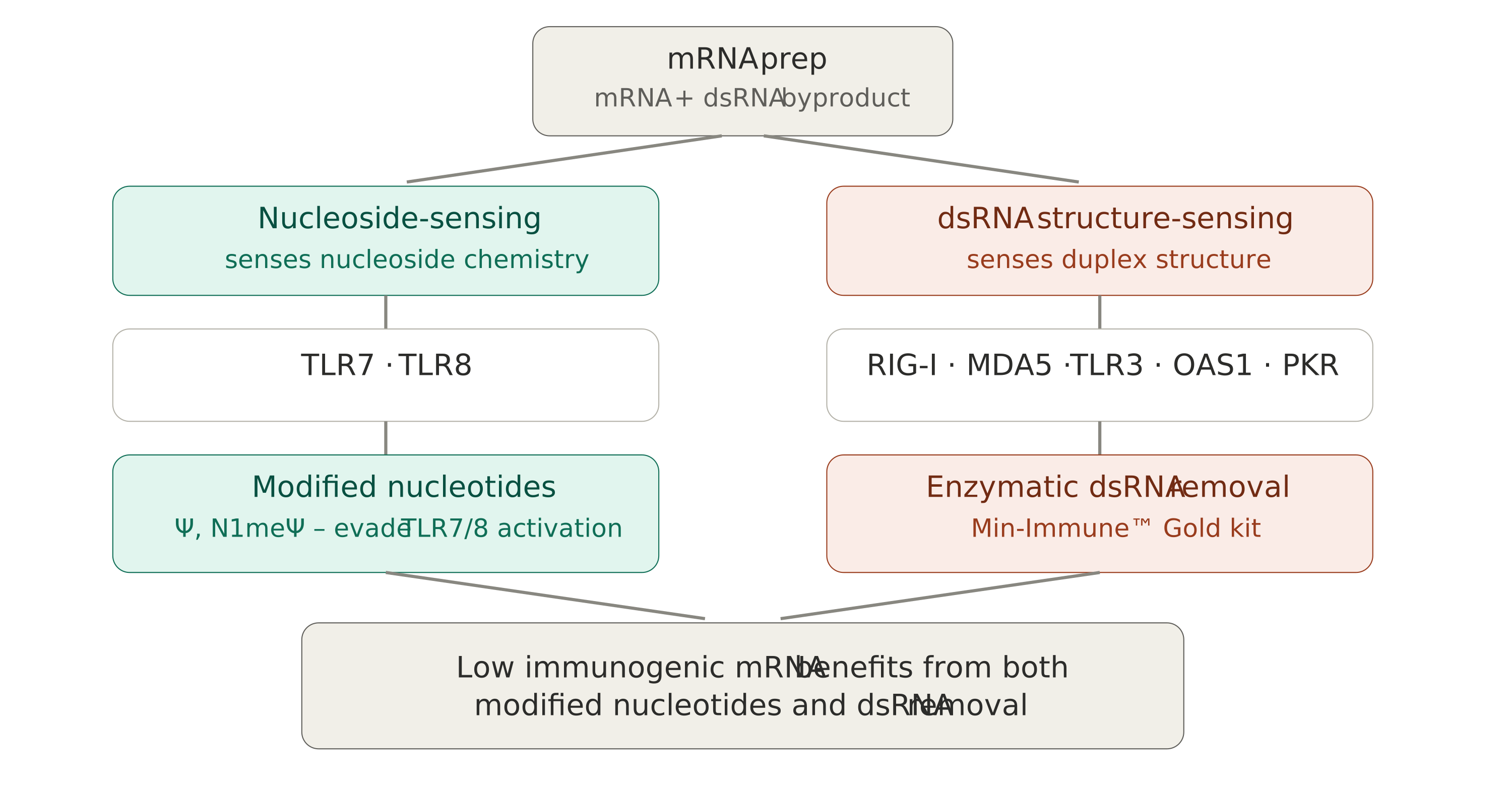
Figure 1. Modified nucleotides quiet the nucleoside-sensing class; the dsRNA structure sensors are addressed only by removing the dsRNA.
We tested this directly. NanoLuc reporter mRNA was synthesized with canonical UTP, pseudouridine, or N1-methyl-pseudouridine, then split into treated and untreated groups using enzymatic dsRNA removal (Min-Immune™ Gold kit). Across three cell lines and two timepoints, canonical UTP mRNA showed luciferase reporter signal gains up to 55-fold after dsRNA removal, with an average ~24-fold improvement across all conditions tested; the exact magnitude depended heavily on how immune-sensitive the cell line was. Ψ containing mRNA gained up to 4.6-fold. N1meΨ-mRNA, which starts from a lower baseline of immune activation, still gained roughly 1.8-fold on top of the modification (2). This pattern isn’t unique to our data: using a different purification method, Karikó and colleagues reported similar data in 2011 — the largest translation gains in unmodified mRNA, and progressively smaller gains as nucleoside modification increased (1).
Why it matters: researchers who’ve already switched to modified nucleotides and are seeing good results might reasonably assume they’ve solved their immunogenicity problem. The sensor data says otherwise. Across eight innate immune genes tested, the receptors most directly responsive to dsRNA structure — RIG-I, TLR3, MDA5 and OAS1 — dropped by 81–86% after enzymatic dsRNA removal, independent of which nucleoside went in (2).
For self-amplifying RNA, the usual fix mostly isn’t available
Self-amplifying RNA constructs retain a viral replicase to drive intracellular replication. The uridine modifications that lower immunogenicity in conventional mRNA — Ψ and N1meΨ — impair that replicase, so they’ve had limited use in saRNA (3). Workarounds are an active research area but aren’t yet routine. For most saRNA workflows that puts the standard nucleoside strategy largely out of reach, and makes dsRNA removal — which is independent of nucleotide chemistry — one of the few practical levers for controlling immunogenicity.
We tested a 9.7-kb VEE-based saRNA construct encoding GFP, comparing untreated material to material treated with the Min-Immune™ Gold kit for 15 or 60 minutes. In THP-1 monocytes, untreated saRNA triggered strong upregulation of dsRNA sensors and cytokines, with IFNB and IL-6 induction reaching roughly 50-fold and greater than 120-fold over control, respectively. Both treatment durations suppressed this response to near-baseline levels, and in HEK293 cells restored functional GFP expression with preserved cell viability (4). The 15-minute treatment performed as well as the 60-minute — worth knowing if transcript clipping from longer treatment times is a concern for your construct.
What this means for your workflow
Modified nucleosides are still worth reaching for first on standard mRNA — they do real, measurable work reducing immune activation. But they cover one class of sensors, not both, and the data above suggests dsRNA removal is worth treating as a separate, additive step rather than assuming nucleoside chemistry already handles it.
Enzymatic removal using RNase III selectively degrades dsRNA while leaving single-stranded RNA intact. Cellulose and reverse-phase HPLC cleanups recover purity but tend to cost you yield; enzymatic removal avoids that, and runs on standard lab equipment with no dedicated chromatography setup.
If you’re troubleshooting inconsistent IVT results, working with a construct where modified nucleosides aren’t an option, or just haven’t checked your dsRNA content in a while, it’s worth a look — more of your workflow’s underperformance than you’d expect may trace back to a byproduct you weren’t testing for.
Full data referenced above, along with our current enzymatic dsRNA removal options, is available below.
References
-
1.
Karikó K., Muramatsu H., Ludwig J., Weissman D. (2011) Generating the optimal mRNA for therapy: HPLC purification eliminates immune activation and improves translation of nucleoside-modified, protein-encoding mRNA. Nucleic Acids Res. 39(21):e142. -
2.
Parker J., Lahr R., Meis R., Meis J. (2026) Enzymatic Elimination of dsRNA Byproducts Enhances Translation and Reduces Innate Immune Activation of mRNA. Presented at ASGCT 2026. -
3.
Quintana V. et al., (2025) Improvement in the potency of a N1-methylpseudouridine-modified self-amplifying RNA through mutations in the RNA-dependent RNA polymerase. J. Biol. Chem. 301(8):110487. -
4.
Meis J., Khanna A. (2026) Min-Immune™ Gold-Mediated dsRNA Depletion Restores Expression and Viability of VEE Self-Amplifying RNA. CELLSCRIPT™ Application Note 006.
